
Traumatic Brain Injury Health Center
Table of Contents
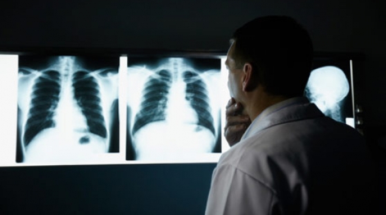
Imaging tests help in determining the diagnosis and prognosis of a TBI patient. Patients with mild to moderate injuries may receive skull and neck X-rays to check for bone fractures or spinal instability. For moderate to severe cases, the imaging test is a computed tomography(CT) scan.
The doctor may order a CT scan or MRI to see pictures of the inside of the brain. These pictures can help the doctor see signs of hematoma or other injuries. Even if none are found, he or she may still decide to keep the patient at the hospital to be watched closely.
In some cases, the doctor may send the patient home with a responsible adult. The doctor may ask the adult to wake the patient up frequently and ask “Where are you?”, “What is your name?”, and “How are you feeling?” to make sure everything is okay.
CT scans may be repeated frequently in the first few days to check if blood clots are getting bigger or if the brain is swelling. MRI scans may be done later to check for more subtle damage that cannot be seen on CT scans. MRIs use strong magnets and radio waves to create detailed images of the inside of the brain. CT scans are more specific for bleeding in and around the brain and can be done faster. This is why CT scans are usually done first.