
Osteoporosis Health Center
Table of Contents
Osteoporosis is usually diagnosed by using a bone mineral density test or a spine CT (computed tomography) scan.
Bone Mineral Density Test
If a patent is at risk for osteoporosis, a doctor may recommend a bone mineral density (BMD) test. This test measures the amount of calcium and other minerals in an area of a bone. This procedure can be done in a few different ways. Most commonly, a dual-energy X-ray absorptiometry (DEXA) scan will be used. DEXA is the most accurate test for osteoporosis and uses low-dose X-rays. The two types of DEXA scans are:
- Central DEXA. A patient lays on a soft table, usually dressed. A scanner passes over their hip and lower spine. It is the best scan to test for the risk of fractures.
- Peripheral DEXA (p-DEXA). This test uses smaller machines that measure the bone density in the wrist, fingers, leg or heel. These machines are commonly found in doctor's offices, pharmacies, shopping centers and health fairs.
A BMD test requires that the patient remain still, but it is painless. A patient should inform a doctor if she is pregnant before getting a BMD test.
The test results will most often be reported as a "T-score" and a "Z- score." A "T-score" shows the patient's bone density compared to that of a younger woman. A "Z-score" is the patient's bone density compared to that of other people of the same age, gender and race. If either score shows a negative number, then the patient has thinner bones than the standard. The further the number is from zero, the greater the patient's risk for a bone fracture.
Spine CT
If a patient has symptoms in the spine, a doctor may recommend a spine CT. A spine CT is a computed tomography scan of the lower spine and surrounding tissues.
During a spine CT, the patient lies on their back on a narrow table that slides into the center of the CT scanner. When the patient is inside the scanner, a machine's X-ray beam rotates around them. During this process, detectors inside the scanner measure the amount of X-rays that make it through the spine. With this information, a computer creates several images, called slices. A doctor evaluates the results.
The patient must lie still to prevent blurred images caused by movement. Patients may also be asked to briefly hold their breath.
Sometimes an iodine-based dye is injected into the patient's vein before the procedure. This is done to highlight specific areas inside the body and create a clearer image. This dye may cause a slight burning sensation, a metallic taste in the mouth or a warm flushing of the body. However, this is normal and the discomfort will go away in a few seconds.
Other Tests
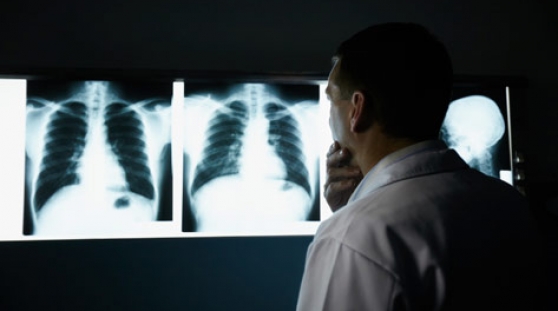
A spine or hip X-ray may show fracture or collapse of the spinal bones in severe cases. However, X-rays of bones are not very accurate in testing for osteoporosis.
Other blood or urine tests may be recommend if it is suspected that the osteoporosis is being caused by a medical condition rather than bone loss caused by aging.