
Brain Cancer Health Center
Table of Contents
If you have symptoms that suggest a brain tumor, your doctor will give you a physical exam and ask about your personal and family health history. You may have one or more of the following tests:
- Neurologic exam: Your doctor checks your vision, hearing, alertness, muscle strength, coordination, and reflexes. Your doctor also examines your eyes to look for swelling caused by a tumor pressing on the nerve that connects the eye and the brain.
- MRI: A large machine with a strong magnet linked to a computer is used to make detailed pictures of areas inside your head. Sometimes a special dye (contrast material) is injected into a blood vessel in your arm or hand to help show differences in the tissues of the brain. The pictures can show abnormal areas, such as a tumor.
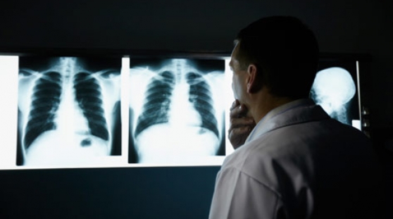
- CT scan: An x-ray machine linked to a computer takes a series of detailed pictures of your head. You may receive contrast material by injection into a blood vessel in your arm or hand. The contrast material makes abnormal areas easier to see. Your doctor may ask for other tests:
- Angiogram: Dye injected into the bloodstream makes blood vessels in the brain show up on an x-ray. If a tumor is present, the x-ray may show the tumor or blood vessels that are feeding into the tumor.
- Spinal tap: Your doctor may remove a sample of cerebrospinal fluid (the fluid that fills the spaces in and around the brain and spinal cord). This procedure is performed with local anesthesia. The doctor uses a long, thin needle to remove fluid from the lower part of the spinal column. A spinal tap takes about 30 minutes. You must lie flat for several hours afterward to keep from getting a headache. A laboratory checks the fluid for cancer cells or other signs of problems.
- Biopsy: The removal of tissue to look for tumor cells is called a biopsy. A pathologist looks at the cells under a microscope to check for abnormal cells. A biopsy can show cancer, tissue changes that may lead to cancer, and other conditions. A biopsy is the only sure way to diagnose a brain tumor, learn what grade it is, and plan treatment.
Surgeons can obtain tissue to look for tumor cells in two ways:
- Biopsy at the same time as treatment: The surgeon takes a tissue sample when you have surgery to remove part or all of the tumor. See the Surgery section.
- Stereotactic biopsy: You may get local or general anesthesia and wear a rigid head frame for this procedure. The surgeon makes a small incision in the scalp and drills a small hole (a burr hole) into the skull. CT or MRI is used to guide the needle through the burr hole to the location of the tumor. The surgeon withdraws a sample of tissue with the needle. A needle biopsy may be used when a tumor is deep inside the brain or in a part of the brain that can't be operated on. However, if the tumor is in the brain stem or certain other areas, the surgeon may not be able to remove tissue from the tumor without harming normal brain tissue. In this case, the doctor uses MRI, CT, or other imaging tests to learn as much as possible about the brain tumor.